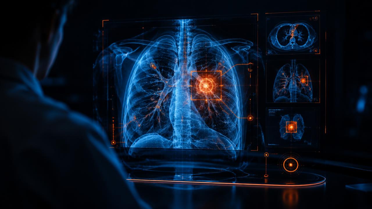
The human body is a universe of complexity. For a clinician, interpreting its signals is a monumental task of pattern recognition. A radiologist might review a single CT scan containing thousands of individual images, searching for a lung nodule just a few millimeters across. A pathologist might spend hours at a microscope, counting abnormal cells on a glass slide to grade a tumor. The work is demanding, the stakes are high, and the volume of data is exploding.
This is the environment where artificial intelligence is making its most profound impact. Not as a futuristic replacement for doctors, but as a tireless, exceptionally skilled collaborator. AI in medical diagnostics is augmenting human expertise, helping practitioners see more, work faster, and deliver more accurate results. It's becoming an indispensable tool in medical imaging, pathology, and even in predicting disease before it fully manifests.
The New Eye in Medical Imaging
The field of radiology is a natural fit for AI. The core task involves identifying visual patterns within medical images, a function at which modern AI, particularly deep learning models, excels. Radiologists face immense pressure, with studies showing significant error rates linked to fatigue and the sheer volume of cases. AI offers a powerful way to mitigate these challenges.
The technology behind this is often a Convolutional Neural Network (CNN). In simple terms, a CNN is a type of algorithm inspired by the human visual cortex. It's trained on vast libraries of labeled medical images, often millions of them, learning to identify the subtle textures, shapes, and anomalies that signify disease. It learns what a healthy lung looks like, what a hairline fracture looks like, and what the faint shadow of an early-stage tumor looks like.
Reading Between the Pixels: From X-rays to MRIs
The applications are already in clinical use and are expanding rapidly.
Finding the Needle in the Haystack: In oncology, AI algorithms can screen chest CT scans and flag suspicious pulmonary nodules for the radiologist's review. Companies like Aidoc and Zebra Medical Vision offer platforms that analyze scans in the background, identifying potential issues from brain bleeds to vertebral fractures. The key benefit is not just spotting something a human might miss, but also triaging the workload. An AI can instantly identify a scan showing a life-threatening condition, like a pulmonary embolism, and push it to the top of the radiologist’s worklist. This prioritization can dramatically shorten the time to treatment.
Preventing Blindness: One of the most successful examples is in ophthalmology. Diabetic retinopathy, a complication of diabetes, is a leading cause of blindness. Early detection and treatment can prevent vision loss, but it requires a trained specialist to examine retinal images. Google and its affiliate Verily developed an AI that can detect diabetic retinopathy from retinal fundus photographs with an accuracy level comparable to board-certified ophthalmologists. This democratizes expertise, allowing screenings to happen in primary care clinics or even mobile vans, with results available in minutes instead of weeks.
Optimizing Stroke Care: In stroke treatment, every second counts. Viz.ai created an FDA-cleared platform that uses AI to analyze brain scans for suspected large vessel occlusion strokes. The system automatically alerts the entire stroke care team, including neurologists and surgeons, on their mobile devices. This can shave critical minutes, or even hours, off the time it takes to get a patient to intervention, directly improving their chances of a good outcome.
Digitizing the Microscope: AI in Pathology
Pathology is the cornerstone of cancer diagnosis. Traditionally, it's a manual and somewhat subjective process. A pathologist prepares a tissue sample, places it on a glass slide, and examines it under a microscope to make a diagnosis and grade the disease. The rise of digital pathology, which involves scanning these glass slides to create high-resolution whole-slide images (WSI), has created the data-rich foundation for AI to thrive.
A single WSI file is enormous, often multiple gigabytes. Manually analyzing it is as daunting as reviewing thousands of CT images. AI provides the computational power to analyze every single cell on that slide.
Counting Cells and Grading Tumors
AI algorithms are being trained to perform tasks that are laborious and prone to inter-observer variability, bringing a new level of objectivity to the field.
Objective Cancer Grading: For diseases like breast and prostate cancer, grading the tumor's aggressiveness is crucial for determining the treatment plan. This often involves counting mitotic figures (dividing cells) or assessing gland formation (the Gleason score in prostate cancer). These can be subjective judgments that vary between pathologists. AI systems, like those developed by PathAI and Paige, can analyze the entire slide, precisely count cells, and provide objective, quantifiable data to support the pathologist's assessment. In 2021, Paige received the first-ever FDA approval for an AI product in digital pathology, a system designed to help pathologists find prostate cancer.
Predicting Molecular Markers: In a fascinating development, AI is learning to predict a tumor's genetic makeup directly from its appearance on a standard H&E slide. Certain genetic mutations, which guide targeted therapies, create subtle changes in cell shape and tissue structure that are invisible to the human eye. An AI, trained on slides with known genetic data, can learn to spot these patterns. This could one day act as a screening tool, helping oncologists decide which patients would benefit most from slower and more expensive genomic sequencing.
For pathologists, this technology isn't a threat. It’s an assistant that handles the tedious work, offers a quantitative second opinion, and uncovers information hidden within the image, allowing them to focus their expertise on the most complex diagnostic challenges.
Beyond the Image: Predictive Analytics and Early Detection
Perhaps the most transformative application of AI in diagnostics isn't about identifying existing disease, but about predicting it. By analyzing massive datasets, including Electronic Health Records (EHRs), lab results, genomic profiles, and even real-time data from wearable devices, machine learning models can identify individuals at high risk for future health problems.
This marks a fundamental shift from reactive to proactive medicine. Instead of waiting for a patient to present with symptoms, clinicians can intervene earlier, often when treatments are more effective and less invasive.
From Reactive to Proactive Healthcare
Predicting Sepsis: Sepsis is a life-threatening response to infection and a leading cause of death in hospitals. Its early signs are subtle and easily missed. Several health systems have implemented AI algorithms that constantly monitor patient data in the EHR, including vital signs, lab results, and medications. The model can detect faint patterns that signal the early onset of sepsis, sending an alert to nurses and doctors hours before a human would typically recognize the crisis. This early warning provides a critical window to begin treatment and save lives.
Forecasting Cardiovascular Events: Traditional models for predicting heart attack or stroke risk, like the Framingham Risk Score, are based on a limited number of variables. AI can build far more sophisticated models. By analyzing hundreds of data points from a patient's EHR, Stanford University researchers developed a deep learning algorithm that was significantly better at predicting cardiovascular events. It learns complex, non-linear relationships between factors like cholesterol, blood pressure, lifestyle, and medical history to create a more personalized and accurate risk profile.
Real-World Implementation: Benefits and Hurdles
The potential of AI in diagnostics is clear, but translating that potential into widespread clinical practice involves overcoming significant obstacles.
The Upside for Clinicians and Patients
The benefits are compelling for everyone involved in the care journey. For clinicians, AI tools promise to reduce the burden of repetitive tasks, which is a major contributor to burnout. They act as a decision support system, providing more data and increasing diagnostic confidence. This frees up their time to focus on what humans do best: communicating with patients, handling ambiguous cases, and exercising clinical judgment.
For patients, the advantages are faster results, which means less time spent waiting anxiously for a diagnosis. It means higher accuracy, reducing the risk of a missed finding or a misdiagnosis. It also means broader access to care. An AI algorithm trained by the world's leading specialists can be deployed in any clinic with the right equipment, bringing top-tier expertise to underserved communities.
The Practical Challenges of Adoption
Putting these tools into practice isn't as simple as installing new software. Several critical challenges must be addressed.
Data Quality and Bias: An AI is a reflection of the data it was trained on. If the training dataset predominantly features one demographic, the algorithm may perform poorly for others. This can unintentionally perpetuate and even worsen existing health disparities. Building fair and equitable AI requires curating diverse, representative, and meticulously labeled datasets.
Integration and Workflow: A brilliant algorithm is useless if it's difficult to use. To be effective, AI tools must integrate seamlessly into the existing clinical workflow and IT infrastructure, such as Picture Archiving and Communication Systems (PACS) and EHRs. A solution that adds extra steps or requires clinicians to log into a separate system is unlikely to be adopted, no matter how accurate it is.
Regulation and Validation: Regulatory bodies like the U.S. Food and Drug Administration (FDA) are grappling with how to evaluate and approve these dynamic systems. The FDA has developed a framework for "Software as a Medical Device" (SaMD) and is creating pathways for algorithms that can learn and adapt over time, but ensuring their safety and efficacy remains a complex, ongoing process.
Cost and Infrastructure: The upfront investment can be substantial. Digital pathology requires expensive whole-slide scanners. Running complex deep learning models requires powerful computing servers. The software itself comes with licensing fees. Demonstrating a clear return on investment is essential for hospital administrators to justify the cost.
AI is not an autonomous physician in a digital box. It is a new class of diagnostic instrument, one that extends the senses and cognitive reach of the clinician, just as the microscope and the MRI did in their own eras. The goal has always been a clearer picture of human health. With AI, that picture is coming into focus faster and with greater detail than ever before. The primary challenge is no longer proving technological capability, but navigating the complex work of integrating it safely, ethically, and effectively into the very human practice of medicine.